
Case study
You are asked to see a patient who was admitted yesterday with a head injury and lacerated scalp. She has a 3-4 year history of increasing falls. On examination, she had retrocollis, lid retraction, an inability to look upwards, and was almost mute. On getting her to stand, she has marked postural instability.
Q1: What is the problem with her eye movements?
Q2: What manoeuvre needs to be done to make the diagnosis?
Q3: What is the neurophysiology of postural instability?
Q4: What is the most likely diagnosis?
Q5: What other clinical signs is she likely to have?
Answers:
Q1: What is the problem with her eye movements?
She has a supranuclear gaze palsy
Supranuclear gaze palsy (SGP) is a complex neurological condition affecting the brain's ability to control eye movements. It primarily impacts the mechanisms that coordinate and execute the movements necessary for the precise tracking of visual targets, smooth pursuit, and rapid shifts of gaze (saccades). This impairment results from a dysfunction in the areas above the nuclei of the cranial nerves that directly control eye movements, hence the term "supranuclear."
Pathophysiology
SGP can arise from various causes, including stroke, brain tumours, neurodegenerative diseases (such as progressive supranuclear palsy, multi-system atrophy, corticobasal ganglionic degeneration), and traumatic brain injuries. The underlying pathology involves damage to or dysfunction of, the brain regions responsible for generating the signals that direct eye movements. The three key structures that control the vertical gaze centre include the rostral interstitial nucleus of the medial longitudinal fasciculus (riMLF), the interstitial nucleus of Cajal (INC), and the posterior commissure (PC). The riMLF is located in the midbrain and helps with vertical and torsional saccades.
Clinical Features
The hallmark of supranuclear gaze palsy is the difficulty or inability to move the eyes vertically, especially in the downward direction. Patients may also experience problems with horizontal gaze, particularly when attempting to look laterally. Initially, these difficulties might be subtle, but they can progressively worsen. Other symptoms may include:
Difficulty in the smooth pursuit of moving objects makes activities like reading or watching moving objects challenging.
Impaired blinking and eyelid control lead to a characteristic "staring" appearance.
Balance and coordination problems, as many regions involved in eye movement, are also implicated in these processes (see below).
Diagnosis and Management
Diagnosing supranuclear gaze palsy requires a comprehensive neurological evaluation, including detailed assessments of eye movements. Imaging studies such as MRI can help identify underlying structural brain abnormalities.
The management is typically directed at the cause of the SGP. There is no cure for SGP, and treatment focuses on managing symptoms and supporting the patient's quality of life. Rehabilitation therapies, such as speech and occupational therapy, can help patients adapt to their symptoms.
Conclusion
Supranuclear gaze palsy is a significant neurological condition that can markedly impair a person's ability to interact with their environment. By affecting the supranuclear pathways that control eye movement, it disrupts the fundamental human experience of visually exploring the world. Despite its challenges, ongoing research and supportive care can help manage its symptoms and improve the quality of life for those affected.
Q2: What manoeuvre needs to be done to make the diagnosis?
To diagnose SGP, one of the key manoeuvres utilised is the ocular motility examination, which assesses the full range of eye movements. Specifically, clinicians often rely on the doll's head manoeuvre to detect supranuclear impairments, known as the oculocephalic and vestibular-ocular reflex (VOR) tests. These manoeuvres help differentiate between supranuclear gaze palsies and other eye movement disorders, such as nuclear or infranuclear palsies.
Doll's Head Maneuver
The doll's head manoeuvre entails moving the patient's head side to side or up and down while observing the eyes' movement. In a healthy individual, the eyes should move in the opposite direction of the head movement, maintaining fixation on a stationary target (this is due to the intact vestibulo-ocular reflex). However, in patients with supranuclear gaze palsy who cannot voluntarily move their eyes towards the direction of the head movement due to the palsy, this manoeuvre might temporarily restore their ability to move their eyes in the said direction. If the eyes correctly move in the opposite direction of the head movement, it suggests that the eye movement muscles and the nerves that innervate them are functioning appropriately, indicating that the problem is above the level of the nuclei (supranuclear).
Vestibulo-ocular Reflex (VOR) Test
Like the doll's head manoeuvre, the VOR test is based on the principle that when the head is turned rapidly to one side, the eyes will move equally and oppositely to maintain fixation on a visual target. This reflex is preserved even in cases of supranuclear gaze palsy because it bypasses the cerebral cortex and directly involves the vestibular system, cranial nerve nuclei, and the muscles controlling eye movement. The preservation of VOR in the presence of impaired voluntary gaze strongly suggests a supranuclear pathology.
Conclusion
These manoeuvres are valuable in diagnosing supranuclear gaze palsy, helping clinicians distinguish it from other types of ocular motor dysfunction. However, a comprehensive evaluation, often including neuroimaging and a detailed neurological examination, is necessary to identify the underlying cause and to plan appropriate management strategies.
Q3: What is the neurophysiology of postural instability?
Postural instability refers to difficulty maintaining an upright posture and balance. This issue is often observed in various neurological disorders, such as Parkinson's disease, and can significantly affect an individual's quality of life. Understanding the neurophysiology behind postural instability involves exploring the complex integration of sensory inputs, central processing, and motor outputs that govern balance and the maintenance of posture.
Key Components
Sensory inputs: three primary sensory systems contribute to postural control:
Visual System: provides information about the environment, orientation, and motion relative to surroundings.
Vestibular System: detects head movements and helps maintain balance during motion.
Somatosensory System: receives information from muscles, joints, and skin about body position and movement (proprioception).
Central processing: The brain integrates the information from the sensory inputs. This integration occurs in several areas:
Cerebellum: coordinates voluntary movements, balance, and posture. It adjusts motor output to make movements smooth and coordinated.
Basal ganglia: involved in the regulation and initiation of movements, as well as muscle tone and posture control.
Brain stem: houses the vestibular nuclei (processing vestibular information) and pathways that mediate the vestibular reflexes essential for balance.
Cortex: contributes to conscious awareness or perception of body positioning and movement.
Motor outputs: signals are sent to the muscles necessary for maintaining posture and balance through descending pathways from the pedunculopontine nucleus and cerebellum.
Pathophysiology of postural instability
Postural instability can result when any part of this intricate system is disrupted. The disruption could be due to:
Sensory impairment: a deficiency in any sensory system reduces the accuracy of the information received, making it difficult to maintain balance.
Central processing problems: disorders affecting the brain regions responsible for integrating sensory information and coordinating motor responses can impair postural stability. This includes the cerebellum and brainstem nuclei, in particular the pedunculopontine nucleus.
Motor systems dysfunction: damage to the motor pathways or muscles can hinder the execution of postural adjustments, contributing to postural instability.
Postural instability often arises from a combination of factors. This patient’s postural righting reflexes were affected. When falling, you would have expected her to extend her limbs to break the fall. The fact that she fell and sustained a head injury leads one to expect this. Therefore, in falling patients who sustain head, facial, and proximal upper limb injuries (e.g., fractured head of humerus), please check for postural instability and loss of righting reflexes. In comparison, patients who sustain distal limb injuries when falling (e.g., fractured writs or Colles fracture) indicate that their righting reflexes are maintained.
Conclusion
The neurophysiology of postural instability involves a complex interaction between sensory information, central processing, and motor responses. Disruptions in this system can lead to difficulties in maintaining an upright posture. Understanding these mechanisms is crucial for diagnosing the condition and developing interventions and therapies to improve balance and reduce the risk of falls in affected individuals.
Q4: What is the most likely diagnosis?
Progressive supranuclear palsy
Q5: What other clinical signs is she likely to have?
Dementia - seen in about one in five cases.
Changes in personality (apathy, a lack of inhibition, anxiety, and a profound state of unease or dissatisfaction)
General slowing of movement and visual symptoms
Slurring of speech
Difficulty swallowing
Poor eyelid function
Contracture of the facial muscles
Retrocollis or backward tilt of the head with stiffening of the neck muscles
Sleep disruption
Urinary incontinence
Constipation
Visual symptoms:
Difficulty reading due to the inability to look downwards and make vertical saccades.
Square-wave jerks
Difficulties with convergence (convergence insufficiency)
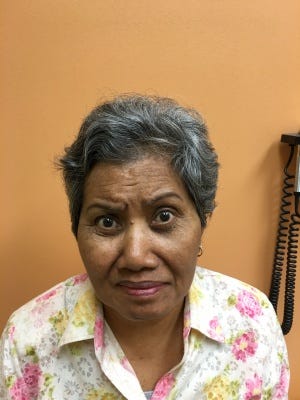
A characteristic facial appearance - procerus sign, with a wide-eyed stare, furrowing of the forehead with a frowning expression, and deepening of other facial creases.
Other signs of Parkinsonism
Twitter / LinkedIn / Medium / MS-Selfie
General Disclaimer: Please note that the opinions expressed here are those of Professor Giovannoni and do not necessarily reflect the positions of Barts and The London School of Medicine and Dentistry nor Barts Health NHS Trust.